Technological Advancements for Early Detection of Breast Cancer
By harnessing AI, breast imaging facilities enhance their capabilities, leading to more effective screenings. This technological advancement is a beacon of hope in our efforts to detect breast cancer early, ultimately saving lives and improving outcomes for countless individuals.
“With the help of AI, radiologists decrease their false positive rates by 37.3% and reduce requested biopsies by 27.8%, while maintaining the same level of sensitivity. This underscores AI’s transformative impact on breast ultrasound diagnosis, enhancing the accuracy, consistency, and efficiency of breast ultrasound diagnosis.”- Nature Communications
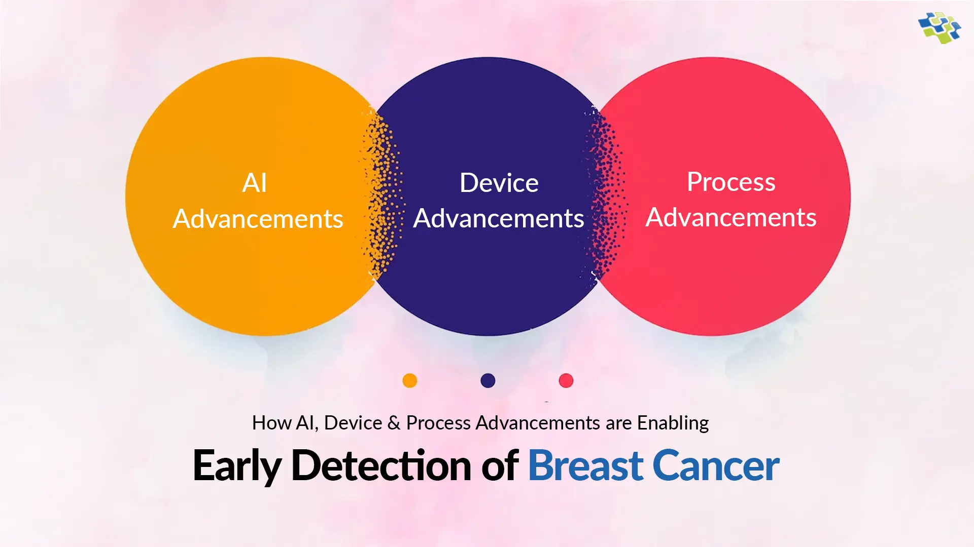
How AI, Device & Process Advancements Enabling Early Detection of Breast Cancer?
AI, Device, and Process advancements are revolutionizing early detection of breast cancer. Together, these innovations enhance efficiency, accuracy, and early intervention, significantly improving breast cancer outcomes. We have included a detailed description of these three advancements below, showing their important contributions to the early detection of breast cancer and their potential to improve healthcare outcomes.
 1. AI Advancements
 1. AI Advancements
AI advancements have significantly improved the early detection of breast cancer. These technologies, especially in ultrasound images, demonstrate accuracy comparable to radiologists. Researchers at NYU Langone Health and the Perlmutter Cancer Center created an advanced AI tool enhancing breast cancer imaging accuracy. Trained on 288,767 ultrasound exams from 143,203 patients, it identifies patterns in images, aiding precise diagnoses. This groundbreaking analysis represents the most extensive of its kind, showcasing significant progress in the early detection of breast cancer.
The Nature Communications journal ‘Artificial Intelligence System Reduces False-Positive Findings in Breast Ultrasound Exams‘ highlights AI’s effectiveness in minimizing erroneous interpretations:
- The paper unveils an AI system reaching radiologist-level precision in breast cancer identification via ultrasound. Trained on 288,767 exams, including 5,442,907 B-mode and Color Doppler images, the AI achieves an impressive AUROC of 0.976 on a test set comprising 44,755 exams, showcasing its remarkable accuracy in diagnosis.
- In a retrospective reader study, the AI outperforms ten board-certified breast radiologists with an AUROC of 0.962, surpassing the radiologists’ average of 0.924±0.02. The AI system reduces false positive rates by 37.3% and biopsy requests by 27.8%, maintaining consistent sensitivity levels. This highlights its superior diagnostic accuracy and efficiency.
Utilizing Artificial Intelligence significantly enhances breast cancer detection, surpassing human capabilities. Radiologists employing AI-assisted screening exhibit superior cancer detection rates compared to their non-assisted counterparts, increasing detection rates by 20% based on studies. AI applications in breast cancer screening predominantly involve object detection (segmentation) and tumor classification, differentiating between benign and malignant cases. The integration of artificial intelligence alleviates radiologists’ workload, enabling more accurate analysis within specified timeframes. A paper titled “Breast Cancer Classification from Ultrasound Images Using Probability-Based Optimal Deep Learning Feature Fusion” introduces an innovative framework employing deep learning and optimal feature fusion for breast cancer classification from ultrasound images, representing a significant advancement in the field.
- Data augmentation expands original datasets to enhance Convolutional Neural Network (CNN) model learning. Utilizing a pre-trained DarkNet-53 model, the output layer is adjusted according to augmented dataset classes for improved performance.
- The adapted model undergoes transfer learning, extracting features from the global average pooling layer for enhanced performance and accuracy.
- Optimal features are chosen via advanced methods: reformed differential evaluation (RDE) and reformed gray wolf (RGW). These features undergo fusion through a novel probability-based serial approach and subsequent classification using machine learning algorithms.
An advanced AI tool, utilizing convolutional neural networks, achieves an 89% accuracy in classifying breast density, significantly contributing to the early detection of breast cancer. Developed by Italian researchers, this software addresses the variability in visual classification, offering transformative potential by enhancing risk assessment. Additionally, AI in ultrasound, coupled with low-cost, handheld probes connected to mobile phones, holds promise for self-administered breast cancer screenings, particularly in rural areas, enhancing early detection efforts and making screening more accessible and affordable for underserved populations. These innovations mark significant strides in the early detection of breast cancer through AI-driven technologies.
2. Device Advancements
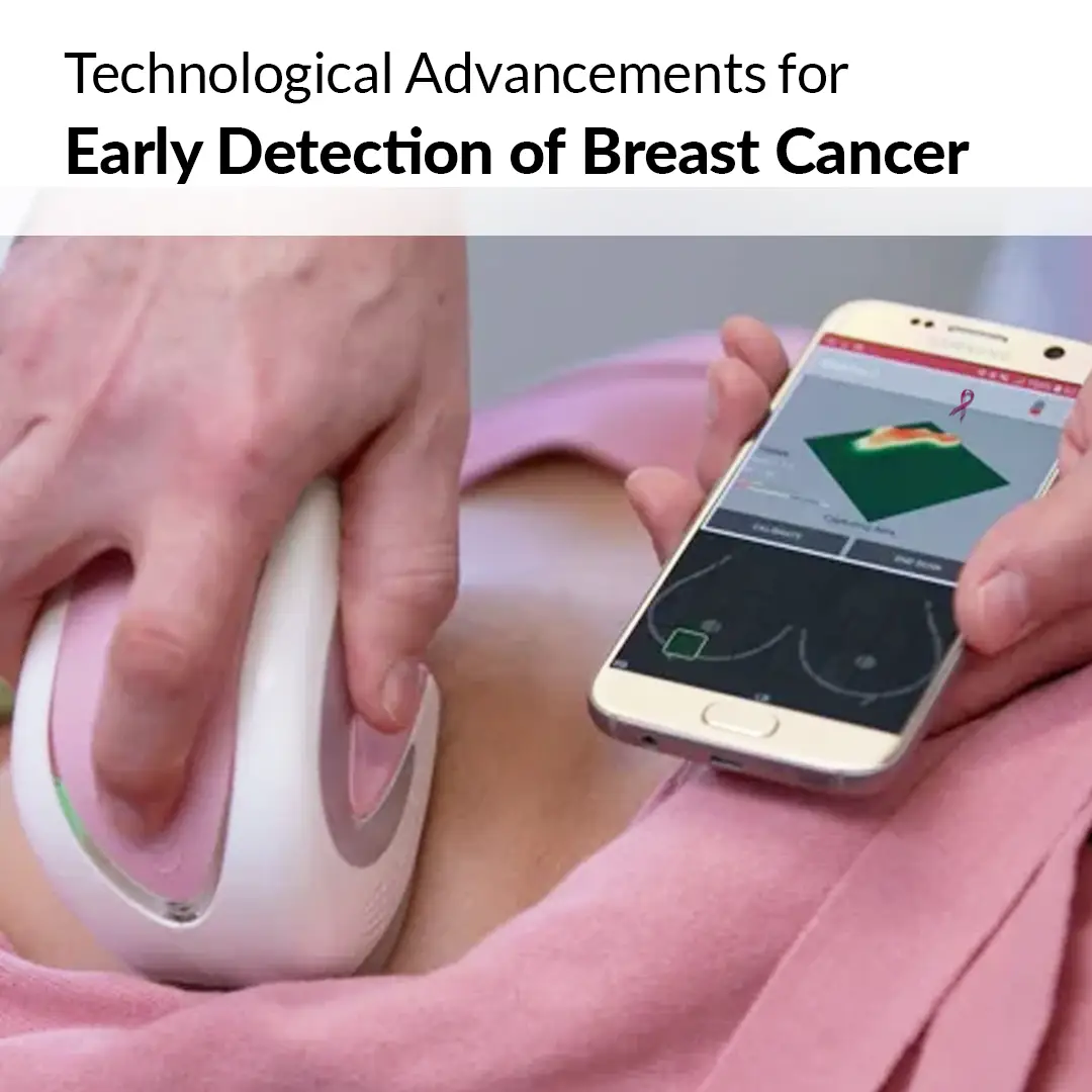
Device advancements, like high-resolution imaging systems and portable ultrasound probes, play a pivotal role in the early detection of breast cancer. These innovations enhance precision, enabling timely screenings and accurate diagnosis, ultimately improving outcomes and saving lives. In the realm of breast cancer detection, ongoing advancements include experimental 3D ultrasound imaging, offering detailed breast images beyond traditional 2D scans. Moreover, portable ultrasound devices compatible with smartphones are emerging, revolutionizing accessibility and early diagnosis methods, and showcasing significant strides in technological evolution for breast cancer screening. Lightweight, handheld ultrasound devices, compatible with smartphones, offer cost-effective and portable imaging solutions. Compared to traditional machines, these innovations facilitate affordable, accessible care, especially benefiting individuals with limited resources. Their portability and affordability mark a significant advancement in healthcare accessibility and early detection efforts.
- The FDA-approved MobiUS device offers versatile imaging capabilities, including cardiac, abdominal, fetal, and pelvic scans. Its approval signifies a significant step in portable, multi-purpose ultrasound technology.
The four key predictions shaping the future of ultrasound technology and patient care. These predictions include:
- Ultrasound Visualization Methods: Ultrasound industry advancements extend beyond traditional 3D and 2D imaging. Innovations focus on refining visualization techniques, aiming for enhanced precision and depth in imaging. These developments signify a significant leap in diagnostic capabilities, ensuring detailed and accurate representations crucial for medical diagnoses.
- Improving Workflow: The medical field is evolving, and ultrasound technology follows suit. Advanced systems feature streamlined interfaces with reduced menus and keystrokes, faster processing, and automated measurements, optimizing workflow efficiency.
- Pocket-Sized Ultrasound Machine: Advancements in pocket-sized ultrasound devices enhance accessibility and portability, democratizing ultrasound technology. These innovations facilitate on-the-go medical imaging, transforming patient care and diagnostics.
- Point-Of-Care Ultrasound Advances: Rapid advancements in point-of-care ultrasound enable swift, precise emergency diagnoses. Enhanced technology accelerates critical decision-making processes, improving patient outcomes in urgent medical situations.
“From Industry to Clinical Practice,” a 2023 Diagnostics journal article, explores breast ultrasound’s evolution from limited grayscale resolution to high-performance, multiparametric modality, highlighting significant technological advancements. The review delves into commercial advancements in breast ultrasound, encompassing microvasculature imaging, high-frequency transducers, elastography, contrast-enhanced ultrasound, 3D ultrasound, and automated techniques. It explores expanded clinical applications, categorizing ultrasound into primary, complementary, and second-look scenarios, emphasizing the evolving role of ultrasound in diverse clinical contexts.
3. Process Advancements
Process advancements in breast cancer detection, like streamlined protocols and automated data analysis, enhance early diagnosis. Improved image interpretation algorithms and efficient reporting enable faster, more accurate assessments, ensuring timely interventions and improved outcomes in the crucial stage of early detection of breast cancer. MIT researchers have developed a revolutionary wearable ultrasound scanner integrated into a bra, allowing frequent at-home monitoring for individuals at high risk of breast cancer. This portable device provides real-time, high-resolution imaging comparable to medical probes. Targeting interval cancers, this portable device aims to increase survival rates to 98% by enabling early detection of smaller tumors, transforming breast cancer diagnosis and patient outcomes. The Conformable Ultrasound Breast Patch (cUSBr-Patch), an innovation from MIT Media Lab, represents a breakthrough in breast cancer detection. This wearable ultrasound patch ensures consistent and reproducible breast imaging, reducing dependence on operator skills and compression techniques. Its standardized approach enhances early detection processes, revolutionizing breast cancer diagnosis.
- The Conformable Ultrasound Breast Patch (cUSBr-Patch) features a honeycomb-shaped design inspired by nature. Equipped with a phased array and user-friendly tracker, it enables extensive, deep, and multi-angle breast imaging.
- In vitro and clinical trials demonstrate the piezoelectric crystal array’s effectiveness, yielding 0.25 mm axial and 1.0 mm lateral resolutions at 30 mm depth. This enables observing small 0.3 cm breast cysts.
- This pioneering research introduces novel ultrasound technology for noninvasive real-time tracking of soft tissue dynamics in breast scanning. The cUSBr-Patch, though tested on a single patient, exhibits accuracy comparable to imaging center ultrasound probes. Its innovation signifies a promising advancement in dynamic soft tissue imaging.
- The cUSBr-Patch was designed for at-risk breast cancer patients to conduct home monitoring. Enabling regular, patient-driven breast tissue surveillance, it enhances proactive healthcare management.
A Radiology study highlighted ultrasound’s effectiveness as a standalone diagnostic method for focal breast complaints. In nearly 2,000 patients, ultrasound alone provided a 90% accurate diagnosis. A meta-analysis showed supplemental ultrasound detected 96% of occult breast cancers missed by mammography and identified 93% of healthy women. Primary ultrasound matched mammography in key metrics but had higher recall rates and detected more invasive cancers. Breast cancer screening and post-treatment imaging surveillance remain suboptimal due to guideline confusion and inadequate attention to dense breasts. Dr. Elizabeth Morris of UC Davis advocated broader MRI utilization and anticipated the integration of artificial intelligence in radiology, foreseeing enhanced accuracy in these practices. Her recommendations underscore the potential of AI and advanced imaging techniques in transforming breast cancer early detection and post-treatment monitoring protocols.
Further Read: How is AI Changing the Future of Precision Medicine?
Conclusion
The synergy of AI, advanced devices, and streamlined processes is revolutionizing the landscape of early detection of breast cancer. AI-driven algorithms enhance accuracy, enabling early diagnosis crucial for improving patient outcomes. The seamless integration of innovative devices, like portable ultrasounds, expands accessibility and empowers proactive monitoring, bridging healthcare disparities. Looking forward, the future of AI in breast cancer detection appears promising. AI systems, continually evolving, will delve deeper into complex data, enhancing pattern recognition and diagnostic precision. As technology advances, AI’s role in the early detection of breast cancer will be pivotal, reshaping the diagnostic paradigm and ultimately saving lives through timely interventions and improved treatment strategies.
Empower your healthcare solutions with NextGen Invent’s cutting-edge AI development services. Harness the power of advanced algorithms and innovative devices to revolutionize the early detection of diseases like breast cancer. Contact us to transform healthcare disparities, ensuring accurate diagnosis and ultimately saving lives.

“In healthcare, the synergy of AI, device, and process innovations illuminates the path to early detection of breast cancer. Embracing cutting-edge technology, redefine possibilities, ensure precision, compassion, and timely intervention, heralding a future where proactive healthcare transforms lives and conquers adversities.”
Deepak Mittal
Founder and CEO
Stay In the Know
Get Latest updates and industry insights every month.