How Is AI in Medical Imaging Improving Diagnostic Accuracy and Workflow Efficiency?


As AI becomes increasingly integrated into medical imaging, healthcare providers must balance its benefits with challenges around data quality, explainability, regulation, and ethics. In this article, we’ll examine how AI is improving imaging workflows while addressing key implementation challenges.

Improving Diagnostic Accuracy but Still Struggling with Imaging Bottlenecks? AI Models Alone Aren’t the Answer. Integrating Intelligence into Clinical Workflows Is What Drives Real Impact
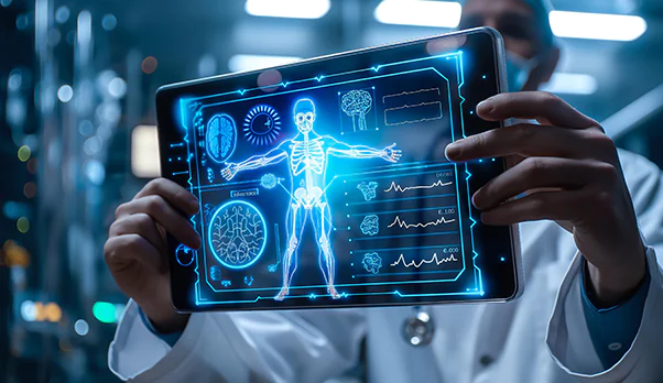
“The fusion of medical imaging and AI has brought about significant advancements in healthcare. From early disease detection to personalized diagnosis and therapy, AI has demonstrated its potential to revolutionize medical practice. By harnessing the power of AI, medical professionals can leverage the wealth of information contained within medical images to provide accurate diagnoses, tailor treatment plans, and improve patient outcomes.” – NCBI
What Is AI in Medical Imaging Workflow Integration?
To put it simply, medical imaging refers to an array of technologies that are used to view the human body to monitor, diagnose, or treat medical disorders. It mostly uses non-invasive visualization methods to help medical professionals detect diseases, chronic issues, and injuries.
AI analysis of medical imaging helps medical practitioners find details or areas of concern that a human may overlook. AI-powered medical imaging can evaluate data points in a medical report to separate signals from noise and diseases from healthy parts.
Medical imaging intelligence has been widely applied to:
- Identify complex patterns and relationships within medical imaging data.
- Assess imaging characteristics using quantitative measurements.
- Recognize imaging modalities and anatomical changes across various stages of treatment.
Challenges Driving the Need for AI in Medical Imaging
Healthcare organizations are keeping up with the substantial backlog of imaging-related requests in the post-pandemic world.
- Healthcare providers are finding it difficult to meet the needs associated with imaging due to the growing number of elderly patients.
- The need for AI clinical decision assistance is growing because of missing or inaccurate diagnoses brought on by human error or subjective interpretations.
- Improved speed and efficiency in imaging analysis and reporting are necessary.
AI medical image analysis helps medical professionals evaluate images, offering a creative solution to these issues. The precision of medical imaging diagnosis, the effectiveness of treatment, and the general standard of patient care are all enhanced by this technology.
Where AI Sits in the Radiology Workflow
AI not only increases accuracy but also streamlines radiological workflows by automating laborious tasks. AI systems can effectively manage repetitive tasks like lesion identification, image segmentation, and the creation of first diagnostic reports. By drastically reducing radiologists’ workloads, this automation improves overall operational efficiency in medical imaging departments and frees them up to concentrate on more complex situations.
- Triage prioritization is one of the most important uses of AI in radiology workflow.
To ensure that critically sick patients receive prompt care, AI systems can evaluate imaging data and indicate urgent situations in real-time, such as identifying cerebral hemorrhages or pulmonary embolisms. This is especially helpful in emergency situations where quick decisions could mean the difference between life and death.
- AI helps minimize delays and ensures the most efficient use of healthcare resources by controlling case prioritization.
- Additionally, AI diagnostic imaging systems can streamline data management by integrating with current Hospital Information Systems (HIS) and Picture Archiving and Communication Systems (PACS).
This medical imaging AI integration makes it possible for improved departmental collaboration, quicker access to imaging results, and more effective case review, all of which are especially helpful in radiology departments with high volume.
Check Our Case Study: AI-Enabled Telepathology Platform Transforming Diagnostic Accuracy
Medical Imaging Modalities Supported by AI
1 AI in Radiology
AI in radiology workflow automates report generation, segmentation, annotation, and triage. It lowers diagnostic error rates and prioritizes urgent cases (such as pneumothorax identification). In retrospective testing, Stanford’s AI model for pneumonia identification fared better than radiologists. AI triage technologies help reduce mammography false positives by 37.3%. These advantages are comparable to advancements in multispectral imaging of burn wounds, where objective data from AI-driven systems decreases subjective visual error and increases consistency.
2. AI in X-Ray Imaging
Ionizing radiation is used by physicians in X-ray imaging to provide two-dimensional images of inside body structures. In this area, AI can examine X-rays to find patterns linked to diseases such as lung nodules, fractures, and specific abnormalities of the chest. Depending on the technology, it can identify questions or categorize results that need more investigation and analysis, giving medical practitioners more details while performing an X-ray scan.
3. AI in MRI
Magnetic resonance imaging (MRI), in comparison to CT and X-ray, uses radiofrequency signals and magnetic fields instead of ionizing radiation to provide detailed images, especially of soft tissues. AI is employed in this discipline to measure, identify essential visual patterns, and segment anatomical structures and lesions. Beyond image analysis, AI is also streamlining MRI workflows by automating complex processes such as implant clearance. At NextGen Invent, we developed ImplantIQ, an Agentic AI-powered platform that extracts implant data, matches device specifications with MRI parameters, and delivers patient-specific safety insights, helping reduce delays and support faster clinical decision-making.
4. AI in Cardiology
To identify structural flaws, perfusion deficiencies, or early indicators of cardiomyopathy, AI models analyze cardiac MRI, CT angiography, and echocardiograms. This facilitates more rapid evaluation of cardiac functions, early identification of atherosclerotic alterations, and more reliable reporting of wall motion, valve function, and EF.
5. AI in CT Imaging
CT creates detailed cross-sectional images of the body by combining X-ray data from various angles, whereas traditional X-rays only give two-dimensional images. AI in CT imaging can segment organs and lesions, identify and describe anomalies, and provide automated measurements on CT scans. It is being used not just for anomaly detection but also for image reconstruction and noise reduction.
Benefits of AI in Medical Imaging Workflow Integration
When we look at medical uses, we can see how AI can be used in a wide range of imaging methods, from new and used ultrasound transducers to X-ray, CT, MRI, mammography, and PET machines.
- Improved Diagnostic Accuracy: Medical imaging intelligence enhances accuracy by spotting trends and anomalies that human eyes might overlook. This lowers the possibility of errors and produces diagnoses that are more precise.
- Personalized Patient Care: AI in medical imaging offers personalized patient care insights based on a patient’s distinct imaging data and additional data points (labs, EMRs, etc.), enabling customized treatment regimens. This increases the effectiveness and patient-specificity of care by enabling doctors to forecast how a disease can progress or react to treatment.
- Faster Image Analysis: AI medical image analysis significantly cuts the amount of time needed to assess medical images. Tasks that used to take hours can now be finished in minutes. Wait periods are shortened, and patient throughput is increased.
- Better Use of Imaging Data: AI can extract quantitative data and patterns that give image evaluation an added layer of information.
- Customized Treatment Plans: AI can analyze vast volumes of patient data, including genetic and medical history. The information gathered can be used to develop individualized treatment programs that are more suited to the needs of each patient.
Check Our Case Study: Clinical Trial Supply Chain Intelligence to Improve Forecasting, Drug Availability & Operational Efficiency
Deep Learning in Medical Imaging Analysis
Deep learning in medical imaging analysis has made strides in image object detection, segmentation, feature extraction, and pattern recognition. Convolutional neural networks (CNNs) have emerged as the leading technique in medical image processing, making it possible to automatically recognize complex imaging patterns and increase diagnostic accuracy.
1. AI in Medical Image Classification
Medical classification and abnormal feature detection are successfully achieved by deep learning models trained on extensive, annotated datasets. A CNN model was tested using a dataset of over 10,000 mammograms. It showed how AI-powered breast cancer screening systems can examine mammograms to check microcalcifications and increase the rates of early cancer diagnosis.
2. AI in Medical Image Segmentation
Lesion organ segmentation and tumor boundary detection are all key applications of AI-driven segmentation techniques. Deep learning algorithms have demonstrated great accuracy in imaging brain tumors, lung lesions, liver cancer, and prostate cancer when applied to MRI and CT image segmentation.
3. AI in Medical Image Reconstruction
AI has improved medical image reconstruction and enhancement in addition to classification and segmentation. While reducing scan time and radiation exposure, AI-driven CT noise reduction and MRI super-resolution reconstruction enhance image quality. For instance, deep learning-based low-dose CT reconstruction methods improve clarity and minimize artifacts, which makes them useful for orthopedic imaging and lung screening.
AI Is Changing Medical Imaging. Is Your Organization Keeping Pace? The Difference Between Adoption and Impact Lies in Building Imaging Workflows That Deliver Measurable Clinical Value
How AI Integrates into Medical Imaging Workflows
Clear roles between clinical and technical teams, dependable data flows, and stable connections to image viewers, radiology information systems, and electronic health record systems are all necessary to turn a model into what radiologists can use daily.
 1. Preparing Imaging Data for AI Analysis
 1. Preparing Imaging Data for AI Analysis
Annotations, segmentation, and imaging quality assurance are primarily clinical in nature. With AI in medical imaging, organizations and radiologists agree on label structures and establish standards that constitute “ground truth”. Once these guidelines and datasets are available, data and IT teams typically collaborate.
- They standardize trial metadata, connect studies to patients, and convert unofficial guidelines into precise, queryable cohort definitions.
This entails establishing inclusion criteria, connecting baseline and follow-up scans, and attaching results in a lung-nodule program so that teams may examine long-term performance and safety.
2. Training, Validating, and Refining AI Models
Teams create monitored pipelines with versioned data, models, and assessment logic on top of these carefully selected datasets.
- In lung nodules, a pipeline can perform “shadow” evaluations before any changes reach the reading room, compare a candidate model to the current one, and routinely re-score new cohorts.
When patients, scanners, or protocols change over time, this helps identify performance drifts.
3. Integrating AI into PACS, RIS, and EHR Workflows
The most beneficial usage of AI outputs is when they are included in already-existing technologies rather than on a stand-alone portal. Without changing the initial images, technical teams usually integrate results using DICOM and HL7® FHIR®.
A structured FHIR Observation or ServiceRequest related to the patient and study can be used to indicate a nodule category or suggested follow-up period for lung screening, with the model version saved for traceability. Radiologists still use their standard viewer; the AI signal is only an extra layer of information that can be heard.
4. Delivering AI Insights to Radiologists and Clinicians
Worklists and summaries are then created by product and radiology teams to align with regular procedures. While a small panel inside the viewer shows interval growth, important metrics, and recommended next steps that can be incorporated into a structured report, a worklist in the same pathway might emphasize patients who are due for follow-up.
The objective is not to replace clinical judgment, but rather to reduce clicks and improve cues.
5. Monitoring Performance and Governing AI Systems
Mature setups keep track of the history of data, code, and models, keep full audit logs, and allow safe rollbacks on containerized infrastructure that is ready for GPUs. Each deployment of an updated lung-nodule model can be associated with a particular dataset slice, evaluation report, approval decision, and timestamp. In the event of a problem, this end-to-end trail helps with root-cause analysis, regulatory inquiries, and internal quality assessment.
Leaders must consider issues, drugs, laboratories, interactions, and results across locations and releases in addition to DICOM before making decisions based on imaging data. By combining non-imaging context with imaging findings, FHIR-native data and analytics solutions enable businesses to compare cohorts, monitor the impact over time, and determine how changes in AI in medical imaging impact care.
Faster Diagnoses. Better Workflow Efficiency. Improved Patient Outcomes. The Question Isn't Whether to Adopt AI in Medical Imaging. It's How Quickly You Can Turn Potential into Results
How AI in Medical Imaging Improves Diagnosis Accuracy
- Early Detection of Diseases: AI algorithms can identify rare and less obvious abnormalities in medical images that are difficult for the human eye to detect. This is essential for early detection of diseases such as cancer, heart disease, and neurological disorders. AI models help in the medical imaging diagnosis of breast cancer by analyzing mammograms and spotting lumps and microcalcifications, which helps with early cancer detection.
- Automated Quantification and Segmentation: AI rapidly and accurately distinguishes the extent, size, volume, and position of organs, tumors, or any other structure in medical images. This helps track medical issues over time. Medical imaging intelligence is used in neurological imaging to diagnose diseases such as multiple sclerosis and Alzheimer’s by analyzing MRI scan data to divide the brain into several regions.
- Reduction in False Positives and False Negatives: Failure to diagnose is a major problem, particularly in cases of cancer. AI increases the reliability of medical imaging diagnosis by reducing the possibility of either overdiagnosing or underdiagnosing a condition. Computer-aided diagnosis in lung CT scans is quite accurate in distinguishing between benign and malignant nodules.
- Pattern Recognition: AI systems excel in identifying correlations and patterns in images. For example, they can detect brain abnormalities, fractures, or lung nodules more quickly and precisely. CNNs, like radiologists, are adept at processing imaging data and reproducing the process of diagnosis.
Challenges of Medical Imaging AI Integration
- Bias in Training Data: Biased forecasts may result from uneven patient population representation.
- Interpretability & Explainability: Models must produce comprehensible results for medical professionals. Clinical adoption can be hampered by deep learning models, which are sometimes referred to as “black boxes,” if their choices are inexplicable.
- Limited Datasets: Due to privacy constraints and annotation costs, high-quality, labeled medical datasets are hard to come by.
- Data Privacy: It’s crucial to protect patient privacy when utilizing and disseminating medical images for model training.
- Variability in Imaging Modalities: Variations in patient anatomy, imaging procedures, and machine settings all contribute to noise.
Regulatory & Legal Considerations
The use of AI in medical imaging workflow integration raises several intricate legal and regulatory concerns. To ensure compliance and safeguard patient interests, developers and healthcare professionals must comprehend this regulatory framework.
The FDA regulation of AI in the US is the first critical component of the regulatory framework. By concentrating on ensuring the efficacy and safety of AI-powered diagnostic tools, the FDA has been actively involved in establishing standards for deep learning in medical imaging. The FDA’s changing strategy for controlling medical imaging intelligence emphasizes the necessity of precise regulatory frameworks for AI applications.
Another important legal factor is data protection. Compliance with data protection legislation, such as HIPAA in the US and the GDPR in Europe, is crucial since AI systems manage enormous volumes of sensitive patient data. Strict regulations on data management, storage, and exchange are required by these rules, ensuring patient data security and privacy.
However, the legal environment surrounding AI in medical imaging is always changing. To keep up with the rapid progress of AI technologies, new legislation is being developed, and old laws are being reinterpreted. As a result, there is a growing demand for well-balanced regulations that protect patient safety and data privacy while promoting innovation.
Future of Medical Imaging with Artificial Intelligence
The future of AI in medical imaging is highly promising, with ongoing advancements expected to enhance diagnostic accuracy, accelerate workflows, and unlock new possibilities for personalized, data-driven patient care.
- AI-Radiologist Collaboration: Medical imaging software solutions complement radiologists’ skills without taking their place. This ensures more trustworthy interpretations, lessens fatigue, and increases diagnostic confidence.
- Automated Diagnostic Workflows: AI in medical imaging diagnosis minimizes manual labor, prioritizes urgent patients, and automates image processing. By increasing accuracy, radiologists can concentrate on intricate clinical choices and procedures.
- Multimodal Healthcare Data: For patient evaluation, medical imaging AI platforms combine data with genetics and clinical records. This unified view makes data-driven healthcare decisions and better diagnosis possible.
- Personalized & Predictive Imaging: AI in medical imaging workflow integration allows forecasting diseases by analyzing patient data. To improve long-term patient outcomes and treatment programs, it is advantageous to employ specialized developers.
Check Our Case Study: From Months to Minutes: AI-Driven Ingredient Intelligence Revolutionizing OTC Drug Discovery
Conclusion: Building a Safe and Scalable Medical Imaging AI Workflow
Artificial intelligence is redefining medical imaging by enabling faster image interpretation, improved diagnostic precision, and more efficient clinical workflows. From detecting subtle abnormalities to supporting treatment planning and operational decision-making, AI continues to enhance the value delivered by medical imaging software solutions across healthcare settings. As adoption grows, organizations that successfully combine technological innovation with clinical expertise will be better positioned to:
- Improve diagnostic accuracy through advanced AI medical image analysis.
- Accelerate reporting and reduce interpretation turnaround times.
- Enhance workflow efficiency across imaging departments.
- Support earlier disease detection and informed clinical decisions.
- Enable more personalized and data-driven patient care.
At NextGen Invent, we help healthcare organizations translate AI potential into measurable clinical and operational outcomes. As a trusted AI radiology PACS software development company, we build intelligent imaging solutions that integrate seamlessly with existing workflows, automate complex processes, and deliver actionable insights. From AI-powered image analysis and workflow optimization to platforms like ImplantIQ that streamline MRI clearance processes, our team develops secure, scalable, and healthcare-ready solutions that help providers improve efficiency, reduce delays, and elevate patient care.
Frequently Asked Questions About AI in Medical Imaging Workflow Integration

“The value of AI in medical imaging emerges not from isolated algorithmic accuracy, but from thoughtful workflow integration. When clinical context, human oversight, interoperability, and continuous validation work together, AI can sharpen diagnostic confidence, reduce avoidable delays, and translate imaging intelligence into better patient outcomes.”
Michael Kaminaka

Chief Growth Officer
Related Blogs

AI Use Cases in Biopharma: Identifying High-Value Opportunities Across the Value Chain
AI in biopharma has gone from luxury to necessity, spurred by unsustainable costs and failure rates. The traditional R&D for any new drug can cost as much as 1 to 2.5 billion dollars and can take anywhere between 10 to 14 years.

How Data Analytics in Pharma is Transforming Drug Discovery, Clinical Trials, and Personalized Medicine
Did you know that 30% of the global data volume comes from the healthcare sector? It is difficult to have an ideal analytics operational model with a large quantity of data.

How AI Predictive Diagnosis Helps Detect Diseases Earlier and Improve Clinical Decision-Making
Clinicians still have too much administrative burden. There are still many important insights buried in fragmented systems. Manual reviews are still leading to diagnostic delays.
Stay In the Know
Get Latest updates and industry insights every month.